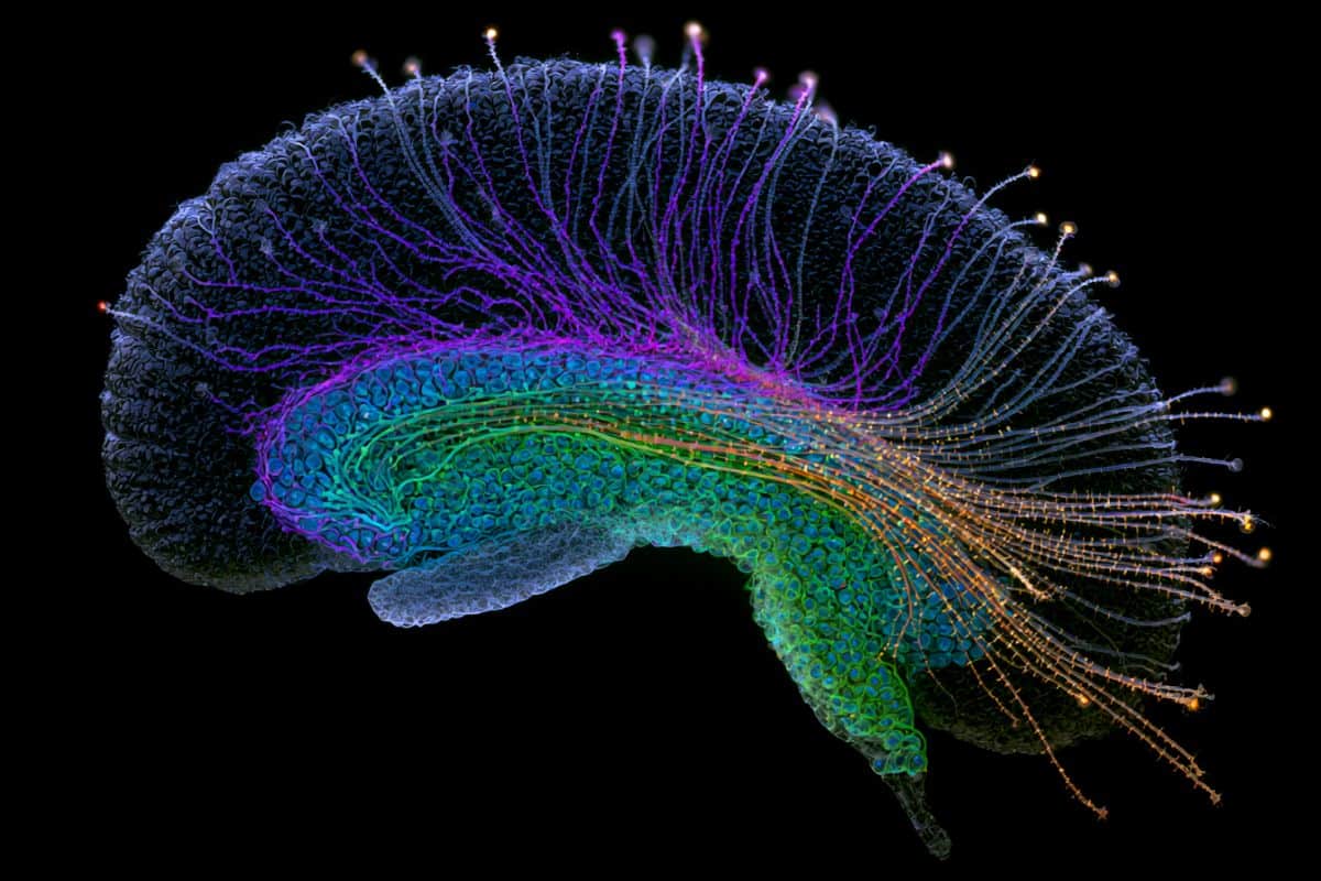
Summary: A new study reveals that the human brain undergoes widespread, coordinated shifts in gene regulation and three-dimensional genome architecture starting in midlife. Examining individual cells across the adult lifespan in the human hippocampus, researchers mapped the structural breakdown of the genome and dynamic system-level remodeling.
A key discovery shows a dramatic shift in the brain’s immune landscape between ages 50 and 75: embryonic-derived microglia decline significantly and are replaced by cells exhibiting peripheral blood immune signatures and elevated inflammatory profiles. Combined with a marked decline in cell populations responsible for maintaining the blood-brain barrier, these genome-level disruptions offer a potential mechanism for why aging remains the primary risk factor for neurodegenerative conditions like Alzheimer’s disease.
Key Facts
- Microglial Replacement in Midlife: Between ages 50 and 75, embryonic-derived microglia decline substantially and are replaced by cells carrying molecular signatures similar to peripheral blood immune cells, displaying elevated pro-inflammatory profiles.
- 3D Genome Architecture Erosion: Across diverse hippocampal cell types, researchers detected a progressive breakdown in the spatial three-dimensional organization of the genome, establishing structural genomic decay as a core hallmark of brain aging.
- Vascular Compromise: The study identified a significant loss of cell populations vital for maintaining blood-brain barrier integrity, increasing vulnerability to circulating toxins and neuroinflammation.
- Multisystem Remodeling: Aging in the human hippocampus is not a passive decay, but a dynamic, coordinated reorganization involving neuronal, vascular, and innate immune networks.
- NIH 4D Nucleome Consortium: This landmark research represents part of a multi-study collection in Science mapping space- and time-dependent genomic architecture across human health and disease.
Source: NYGC
Researchers have discovered that the human brain undergoes widespread age-related changes in genome regulation beginning in midlife that may help explain why aging is the greatest risk factor for neurodegenerative diseases such as Alzheimer’s.
In a new study published in Science, researchers used cutting-edge single-cell technologies to profile gene regulation and three-dimensional genome architecture in individual cells from the human hippocampus, the brain region critical for learning and memory.

By examining brain samples across the adult lifespan, the team generated one of the most comprehensive views to date of how genome regulation changes during brain aging.
One of the study’s most striking findings was a dramatic shift in microglia, the brain’s immune cells, between approximately ages 50 and 75. Microglia that originate during embryonic development declined substantially and were replaced by cells with molecular signatures that resemble immune cells from the blood. This finding challenges the longstanding view that microglia established during embryonic development persist throughout the human lifespan.
These replacement microglia-like cells exhibited elevated inflammatory signatures, suggesting they may contribute to chronic neuroinflammation in the aging human brain. The scientists also observed a substantial decline in cell populations important for maintaining the blood-brain barrier, which protects the brain from harmful substances in the bloodstream.
“Microglia are critical for maintaining brain homeostasis,” said Bing Ren, PhD, a corresponding author of the study, Scientific Director and CEO of the New York Genome Center, Professor of Genetics and Development, Biochemistry and Molecular Biophysics, and Systems Biology at Columbia University, and Associate Director in the Vagelos Institute for Basic Biomedical Science (Vagelos Institute) in VP&S, Columbia University,
“When these cells fail to perform their housekeeping duties, toxic materials accumulate that can trigger inflammatory processes that may contribute to neurodegenerative diseases.”
Across multiple brain cell types, researchers also observed a global erosion of three-dimensional genome architecture, suggesting that structural breakdown of the genome may be a fundamental hallmark of brain aging. “This work represents a major step forward in understanding how aging reshapes the human genome in brain cells,” said Nathan Zemke, Director of Single-cell Genomics at the Center for Epigenomics at UC San Diego. “These findings demonstrate a critical need for studying gene regulation and genome organization to gain a mechanistic understanding of the aging process.”
“Importantly, this study reveals that aging is not simply a gradual decline, but involves coordinated and dynamic remodeling of immune, vascular, and neuronal systems. These findings open the door to identifying new therapeutic targets aimed at preserving circuit integrity and brain function across the lifespan,” said Xiangmin Xu, PhD, Chancellor’s Professor and Director of the Center for Neural Circuit Mapping at the University of California, Irvine, and a co-corresponding author of the study.
This work is one of six papers published in Science as part of the National Institutes of Health’s 4D Nucleome (4DN) Common Fund program, a decade-long initiative designed to map the dynamic architecture of the genome in space and time.
The 4D Nucleome program brought together interdisciplinary teams from across the country between 2015 and 2025 to better understand how the genome’s spatial organization influences biological processes. In addition to this study, Dr. Ren also contributed as a co-corresponding author or co-author on three additional Science papers exploring genome architecture across diverse cell types and timescales.
Collectively, these studies provide a foundational resource for the scientific community and open new avenues for understanding how disruptions in genome organization contribute to development, aging, and disease—including neurodegenerative disease.
Key Questions Answered:
A: Between ages 50 and 75, resident microglia established during embryonic development decline sharply. They are replaced by microglial-like cells that mirror blood-derived immune profiles, which carry higher pro-inflammatory gene signatures and contribute to chronic brain inflammation.
A: 3D genome architecture refers to how DNA is folded and spatially arranged within the cell nucleus to control gene expression. Its structural breakdown across aging hippocampal cells disrupts regular gene regulation, leading to cellular dysfunction and reduced homeostatic maintenance.
A: Rather than viewing brain aging as a slow, uniform deterioration, this study shows that aging triggers dynamic, coordinated shifts in vascular, immune, and neuronal systems starting in midlife. Targeting these specific epigenomic and structural changes could yield interventions to preserve brain function before neurodegeneration takes hold.
Editorial Notes:
- This article was edited by a Neuroscience News editor.
- Journal paper reviewed in full.
- Additional context added by our staff.
About this brain aging and genetics research news
Author: Jessica Leitner
Source: New York Genome Center
Contact: Jessica Leitner – New York Genome Center
Image: The image is credited to Neuroscience News
Original Research: Open access.
“New Study Revises Understanding of Brain Immune Cells During Human Aging” by Nathan R. Zemke, Seoyeon Lee, Sainath Mamde, Bing Yang, Nicole Berchtold, B. Maximiliano Garduño, Hannah S. Indralingam, Weronika M. Bartosik, Pik Ki Lau, Keyi Dong, Emily Hsu, Amanda Yang, Yasmine Tani, Chumo Chen, Qiurui Zeng, Varun Ajith, Liqi Tong, Chanrung Seng, Daofeng Li, Ting Wang, Jingtian Zhou, Joseph R. Ecker, Christopher K. Glass, Carl W. Cotman, Xiangmin Xu, Bing Ren. Science
DOI:10.1126/science.adt8307
Abstract
New Study Revises Understanding of Brain Immune Cells During Human Aging
INTRODUCTION
Aging is the strongest risk factor for neurodegenerative disease and is accompanied by memory decline and chronic inflammation in the brain. The hippocampus, a region essential for learning and memory, is particularly vulnerable to aging. Prior studies have identified age-related shifts in gene expression, including increased inflammatory signaling and reduced synaptic function, implicating transcriptional dysregulation in brain aging. However, gene expression alone provides an incomplete view.
It remains unclear how epigenetic regulation and higher-order genome organization change across cell types and how these changes drive hippocampal dysfunction. Defining these molecular alterations is critical for understanding cognitive decline and disease susceptibility.
RATIONALE
To address this, we profiled gene expression and multiple layers of epigenetic regulation, including chromatin accessibility, DNA methylation, and three-dimensional (3D) genome organization at single-cell resolution across the adult lifespan. This integrated multiomic approach enables direct assessment of how epigenetic and structural genome changes shape transcriptional programs in aging brain cells, and reveals coordinated, cell type–specific mechanisms not captured by single-modality analyses.
RESULTS
Aging is associated with coordinated and often nonlinear changes in gene regulation across cell types, with a major transition occurring around midlife. A notable finding was a remodeling of the brain’s immune cell landscape. Microglia underwent a nonlinear transition in which embryonically derived, brain-resident microglial cells were progressively replaced by a population with epigenetic features resembling blood-circulating monocytes. This transition was not readily detectable using gene expression alone but was revealed by DNA methylation, which preserves cellular lineage. These monocyte-like microglia exhibited epigenetic, transcriptional, and 3D genome features associated with proinflammatory programs, suggesting a potential driver of age-related neuroinflammation.
In parallel, there was a marked decline in astrocytes, including those that support synaptic signaling and maintain the blood-brain barrier. This loss was accompanied by reduced expression of genes involved in mitochondrial energy production and increased cellular stress signatures, indicating metabolic dysfunction as a potential contributor to astrocyte attrition.
At the genome architecture level, aging was marked with a global weakening of 3D genome organization across cell types, including reduced integrity of topologically associating domains and increased trans chromosomal interactions. These structural changes coincided with epigenetic alterations and shifts in gene expression, reflecting widespread rewiring of regulatory programs.
CONCLUSION
Aging reprograms both the cellular composition and regulatory architecture of the human hippocampus. Replacement of embryonically derived microglia with a proinflammatory, monocyte-like population, together with astrocyte loss and global alterations of genome organization, defines key hallmarks of brain aging. These coordinated changes provide a mechanistic framework for age-related memory decline and increased vulnerability to neurodegenerative disease.