Summary: Researchers demonstrated that the success of anti-CTLA-4 immunotherapy against brain tumors does not depend on local T cells alone. Instead, it requires an intensive B-cell and antibody response initiated far away from the tumor, deep within the neck’s lymphatic system.
By unmasking how anti-CTLA-4 triggers the generation of cancer-targeted IgG antibodies within the deep cervical lymph nodes, this discovery rewrites the foundational operating laws of neuro-oncology and provides a brilliant new blueprint to treat intractable brain cancers.
Key Facts
- Shifting the T-Cell Dogma: Conventional oncology has long relegated B cells to the simple production of antibodies against common virus infections or vaccines, while treating T cells as the exclusive shock troops needed to fight cancer. Professor Lee’s lab directly challenged this view by demonstrating that in mouse glioma models, the therapeutic effects of anti-CTLA-4, including dramatic tumor mass reduction and prolonged long-term survival, were completely erased in models lacking B cells, establishing B cells as the absolute gatekeeper of checkpoint efficacy.
- The Deep Cervical Lymph Node Hub: The researchers tracked the primary site of this vital immune response and discovered it does not occur inside the brain tissue itself. Instead, the response takes place distally within the deep cervical lymph nodes, the drainage basins located deep inside the tissue of the neck that filter cerebrospinal fluid leaving the skull.
- The Germinal Center Activation Loop: Following anti-CTLA-4 treatment, the team documented a massive surge of germinal center B cells working in tandem with T follicular helper cells inside these neck lymph nodes. This coordinated cellular machinery triggered an immediate spike in Immunoglobulin G (IgG)—the primary class of high-affinity antibodies.
- Macrophage Recruitment & Direct Opsonization: These newly minted IgG antibodies rapidly migrate from the neck into the brain, where they bind securely to the surfaces of the invading glioma cells. This binding acts like a highly bright digital flare, allowing local macrophages (the immune system’s primary cleanup cells) to easily recognize, engulf, and destroy the cancer cells via enhanced phagocytosis.
- Direct In Vivo Fluorescence Visual Validation: To prove this cellular cleanup loop was happening inside the living tissue, the KAIST team engineered a specialized dual-reporter glioma model. By expressing the red fluorescent protein mCherry alongside the green fluorescent protein EGFP, the team used high-resolution intravital imaging to directly visualize tumor-infiltrating phagocytes actively eating the glowing glioma cells following treatment.
- Expanding the Framework of Cancer Immunotherapy: This study provides the world’s first functional proof that B-cell immune pathways are the deciding factor in determining the success of brain tumor treatments. It completely redefines the landscape of cancer medicine by showing that the survival outcome of an intracranial tumor is heavily governed by systemic immune centers operating completely outside the skull.
Source: KAIST
Researchers have uncovered a clue to why immune checkpoint inhibitors—cancer therapies that release the immune “brakes” exploited by tumors to evade attack—show limited efficacy in some brain tumors.
A KAIST research team found that B cell and antibody responses initiated in tumor-draining lymph nodes, rather than T cells alone, are critical to the antitumor effects of anti-CTLA-4 therapy, opening a new avenue for treating intractable brain tumors.

KAIST (President Choongsik Bae) announced on the 19th of July that a research team led by Professor Heung Kyu Lee from the Department of Biological Sciences has identified a previously unrecognized immune mechanism through which anti-CTLA-4, a type of immune checkpoint inhibitor, promotes B-cell responses in tumor-draining lymph nodes, thereby helping the immune system attack brain tumors.
Glioblastoma is one of the most aggressive malignant brain tumors, with frequent recurrence and a poor prognosis even after surgery and radiation therapy. Immune checkpoint inhibitors, which restore the ability of immune cells to attack cancer cells, have produced substantial therapeutic benefits in various cancers. However, their effectiveness in glioblastoma has remained limited because of the highly immunosuppressive environment surrounding the tumor.
Researchers have traditionally regarded T cells—immune cells that can directly attack cancer cells—as the primary target of immune checkpoint inhibitors. B cells, meanwhile, are well known for producing antibodies following infection or vaccination, but their role in brain tumor immunotherapy has remained largely unexplored. The research team therefore investigated whether anti-CTLA-4 could influence B-cell responses as well as T-cell responses.
The findings challenged the prevailing T-cell-centered view. In mouse glioma models, anti-CTLA-4 treatment reduced tumor burden and significantly prolonged survival. However, these therapeutic effects were largely lost in mice lacking B cells, demonstrating that B cells are required for the efficacy of anti-CTLA-4 treatment in these models.
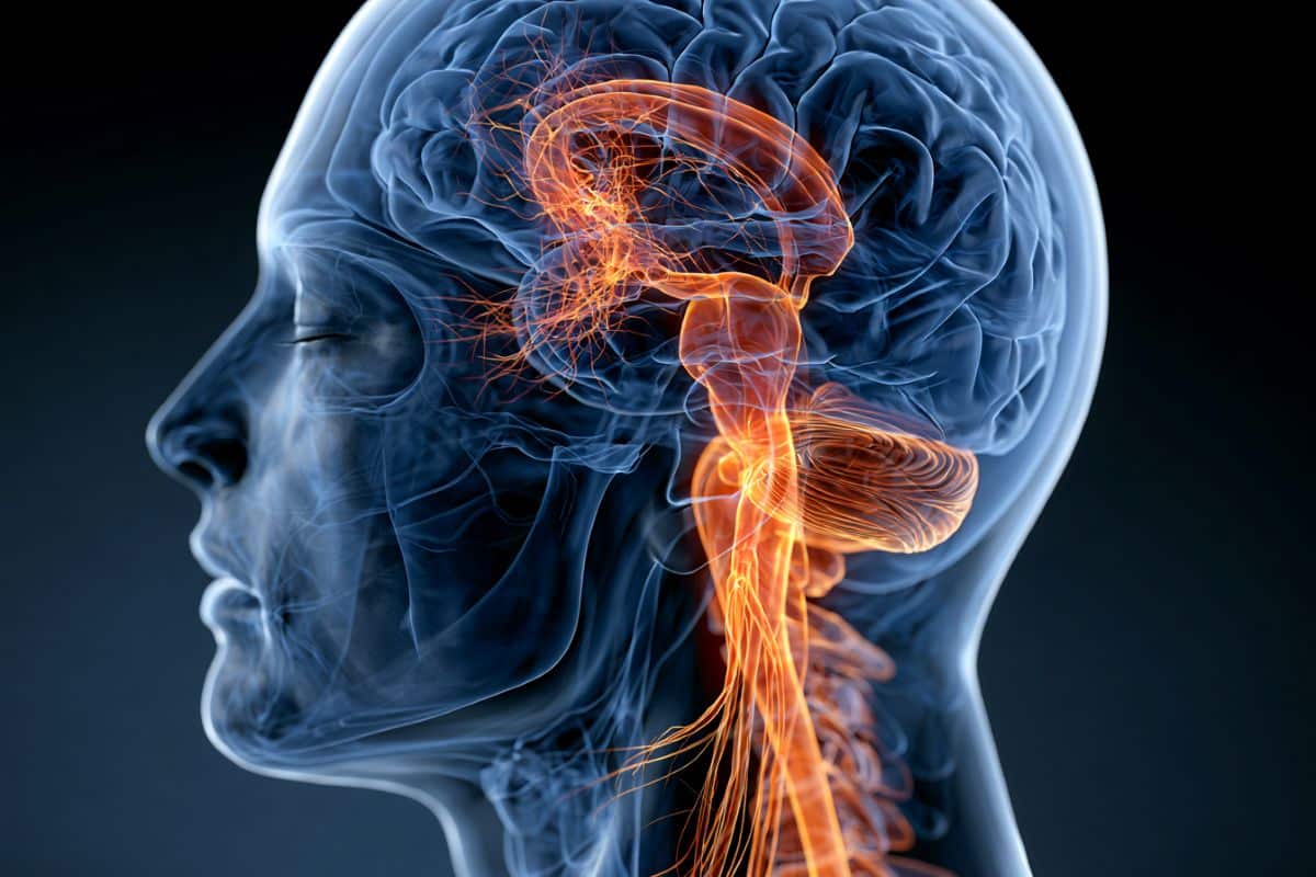
The team also identified where B cells played their key role. Rather than being prominent in the brain, where the tumor cells were located, the response increased markedly in the Deep Cervical Lymph Nodes, which are located deep in the neck and receive lymphatic drainage from the brain.
In particular, germinal center B cells and T follicular helper cells, both of which are important for antibody formation, increased together in these lymph nodes. This was accompanied by an increase in immunoglobulin G, or IgG, responses. IgG is a major class of antibody that can recognize cancer cells as targets and help immune cells eliminate them.
The resulting IgG antibodies bound to the surface of glioma cells, helping macrophages—immune cells that engulf foreign substances and cancer cells—remove the tumor cells more effectively.
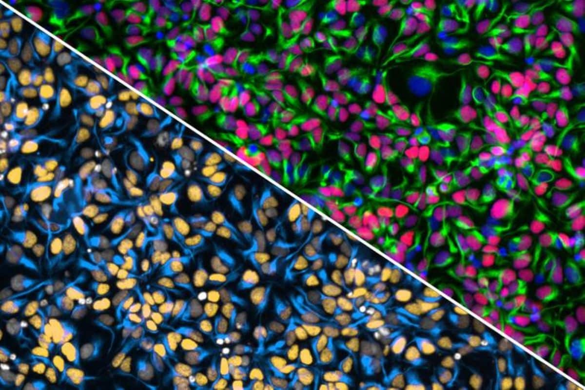
The research team also examined this process directly in vivo. Using a specialized dual-reporter glioma model expressing the red fluorescent protein mCherry and the green fluorescent protein EGFP, the researchers successfully visualized tumor-infiltrating phagocytes actively engulfing glioma cells following anti-CTLA-4 treatment.
The study provides the first functional evidence that B-cell immune responses—previously known mainly for their roles in infection and vaccination—can be a key factor determining the effectiveness of immunotherapy for hard-to-treat brain tumors. It also expands the conventional T-cell-centered framework of cancer immunotherapy by showing that treatment efficacy can be strongly shaped by immune responses originating not only within the tumor, but also in tumor-draining lymph nodes outside it.
Yumin Kim, a postdoctoral researcher in the KAIST Department of Biological Sciences, served as the first author of the study, with Professor Heung Kyu Lee serving as the corresponding author. Professor Ji Eun Oh from the KAIST Graduate School of Medical Science and Engineering also contributed to the research. The findings were published on July 10 in Science Immunology.
Authors: Yumin Kim, In Kang, Byeong Hoon Kang, Won Hyung Park, Chae Won Kim, Hyun-Jin Kim, Jeongwoo La, Myoung Seung Kwon, Sang Hee Park, Seo Hyeon Im, Hyeon Cheol Kim, Keun Bon Ku, Minji Kim, and Ji Eun Oh from KAIST, with Heung Kyu Lee from KAIST as the corresponding author.
Funding: This research was supported by National Research Foundation of Korea grants RS-2023-NR077244 (to H.K.L.), RS-2024-00439735 (to H.K.L.), RS-2024-00411928 (to Y.K.), RS-2025-00517107 (to J.E.O.), and RS-2026-25509011 (to H.K.L.). This study was also supported by Samsung Science and Technology Foundation grants SSTF-BA1902-05 (to H.K.L.) and SSTF-BA2201-11 (to J.E.O.).
Key Questions Answered:
A: Glioblastomas are notoriously brilliant at cloaking themselves from the immune system. They construct an incredibly hostile, highly immunosuppressive local environment within the brain—often referred to as an immunologically “cold” tumor. The moment a killer T cell tries to infiltrate the tumor zone, it is hit with a wall of chemical signals that put its internal brakes on, turning it completely numb. B cells, however, operate like a specialized munitions factory located safely outside the primary war zone. By staying out in the deep cervical lymph nodes of the neck, these B cells are completely insulated from the tumor’s suppression shield. They can safely analyze the cancer’s signature, manufacture millions of high-powered IgG antibodies, and send these homing missiles up into the brain to paint the cancer cells for destruction.
A: Think of your deep cervical lymph nodes like the primary wastewater treatment facilities for your central nervous system. These nodes sit deep along the muscles of your neck. For a long time, traditional medicine believed the brain was completely cut off from the body’s lymphatic system. We now know that the cerebrospinal fluid that bathes your brain constantly drains down out of the skull through tiny channels, heading straight into these specific neck lymph nodes. Because they receive the direct structural runoff from the brain, these lymph nodes act as the ultimate security checkpoint. The moment cells or fluid from an active glioma drain down into the neck, the deep cervical nodes are the very first places where the immune system catches sight of the disease.
A: To definitively prove that the antibodies were successfully calling in reinforcement cells to eat the cancer, the KAIST team built a beautiful visual tracking system inside the living models. They engineered the glioma cells to express two distinct fluorescent tags: a red protein (mCherry) and a green protein (EGFP). When an immune cleanup cell (a macrophage) responds to the IgG antibody flags and engulfs a cancer cell, the highly sensitive dual-fluorescence system changes its light emission under a specialized microscope. This allowed the scientists to watch the exact moment the immune cells surrounded, ingested, and digested the malignant brain tumor tissue in real time, turning theoretical data into concrete, undeniable visual proof.
Editorial Notes:
- This article was edited by a Neuroscience News editor.
- Journal paper reviewed in full.
- Additional context added by our staff.
About this brain cancer research news
Author: Jeonga LEE
Source: KAIST
Contact: Jeonga LEE – KAIST
Image: The image is credited to Neuroscience News
Original Research: Open access.
“The efficacy of immunotherapy in glioma requires distal B-cell responses in tumor-draining lymph nodes” by Yumin Kim, In Kang, Byeong Hoon Kang, Won Hyung Park, Chae Won Kim, Hyun-Jin Kim, Jeongwoo La, Myoung Seung Kwon, Sang Hee Park, Seo Hyeon Im, Hyeon Cheol Kim, Keun Bon Ku, Minji Kim, and Ji Eun Oh. Science Immunology
DOI:10.1126/sciimmunol.adz2494
Abstract
The efficacy of immunotherapy in glioma requires distal B-cell responses in tumor-draining lymph nodes
Brain tumors represent a unique challenge for cancer immunotherapies because of their location in an immune privileged site. However, the brain tumor immune microenvironment is dictated more by tumor type than the location within the brain per se.
This feature is reflected by the higher immunogenicity and response to immunotherapies of metastatic brain tumors compared with primary brain tumors. Immunotherapies for brain tumors aim at inducing and boosting tumor T cell responses using vaccines, immune checkpoint inhibitors, or adoptive T cell therapies.
A fundamental challenge in the field is how such brain tumor–targeting T cells gain access to brain tumors and maintain their function despite a hostile immunosuppressive microenvironment.
Here, we review current knowledge of the cellular and molecular determinants of the antigenicity of brain tumors and the immunosuppressive brain tumor microenvironment.
Expanding and exploiting this knowledge will provide the key for effective combinatorial therapies.