Summary: In 1856, while examining a brain mistakenly believed to belong to the legendary mathematician Carl Friedrich Gauss, Göttingen anatomist Rudolf Wagner documented a bizarre structural quirk: a physical, surface-level connection bridging the central fissure, the deep groove separating the frontal and parietal lobes.
Twenty years later, Viennese anatomist Richard Heschl launched a massive post-mortem audit of 1,087 brains to hunt for this elusive “bridge.” He discovered it in just six cases (0.6%) and hypothesized that it was actually a normal, deep-lying brain fold (a “deep winding”) that occasionally grew unusually high, breaking through to the brain’s surface.
Nearly 150 years later, an international team successfully replicated Heschl’s historic findings using ultra-high-resolution magnetic resonance imaging (MRI).
Analyzing 1,112 healthy adults from the Human Connectome Project, the team not only validated Heschl’s structural theories with precision but also uncovered a brand-new twin-based clue that suggests this rare anatomical signature is shaped by environmental forces in the womb, rather than genetics.
Key Facts
- The 2013 Gauss Swap Discovery: The historical brain that first exposed this rare cortical bridge belonged to a physician named C. H. Fuchs. It had been accidentally swapped with the brain of mathematician C. F. Gauss in the 1860s—a mix-up discovered by Dr. Renate Schweizer in 2013 precisely because of Fuchs’s unique “bridge.”
- Heschl’s Modern Foresight: In 1876, Richard Heschl pioneered a modern “statistical approach” to neuroanatomy by manually auditing over 1,000 post-mortem brains within a single year to track normal vs. rare structural variations.
- MRI Blueprint Validation: Using advanced 3D surface reconstructions of modern MRI data, the researchers found 9 cases of the bridge out of 1,112 subjects, a prevalence of 0.8%, matching Heschl’s original 0.6% calculation.
- The Twin Enigma: The modern study included a heavy subpopulation of twins, revealing that the bridge appeared far more frequently in twins (1.8% of fraternal twins and 1.1% of identical twins).
- Womb Environment Driver: Because the cortical bridge typically appeared in only one twin out of a pair, researchers concluded that this physical landmark is not genetically determined, but is instead sculpted by micro-environmental stressors or space constraints inside the womb.
- Zero Functional Impact: Dr. Renate Schweizer emphasizes that the bridged central fissure is an elegant anatomical variant, not a pathological anomaly or birth defect. It provides no known cognitive advantages, downfalls, or physical changes in human ability.
Source: DPZ
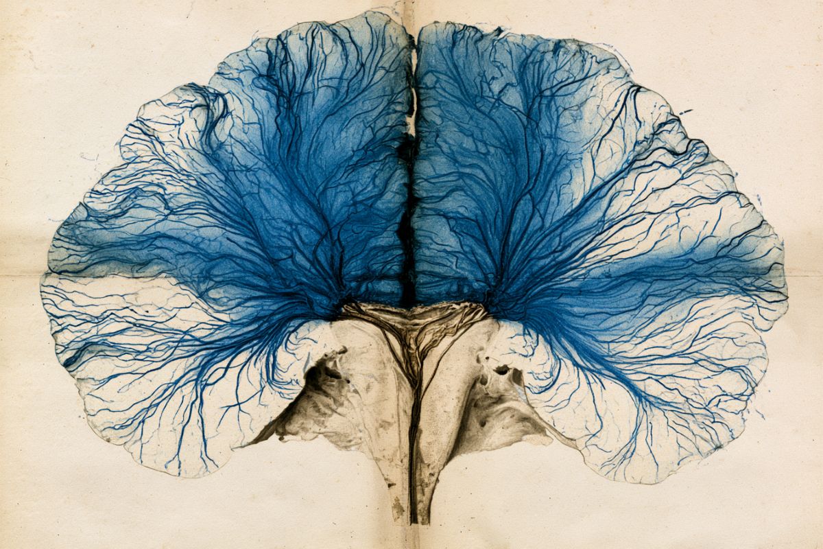
When the Göttingen anatomist Rudolf Wagner examined a brain in 1856 that was later mistakenly believed to be that of the famous mathematician Gauss, he noticed an unknown peculiarity: a connection above the central fissure — which separates two parts of the brain — that he described as a “bridge.”
Twenty years later, in 1876, the Viennese anatomist Richard Heschl searched more than 1,000 brains for further instances of this “bridge” and was able to identify it in six of them. He also measured an ordinary brain winding at the same location, situated at the base of the fissure, as he suspected a connection between this “deep winding” and the “bridge”: if the deep-lying winding occurred at varying heights, the “bridge” should appear at the same location, but at the brain’s surface.
The data confirmed his hypothesis. 150 years later, researchers from the German Primate Center – Leibniz Institute for Primate Research (DPZ) and the University of Göttingen, led by Renate Schweizer, a scientist in the Department of Functional Imaging at the DPZ, have now replicated Heschl’s findings.
For their work, Renate Schweizer, together with her co-authors Anna M. Müllen and Julius Stropel, has been awarded the Replication Prize of the Organization for Human Brain Mapping. The article on which the prize is based was published in 2025 in the journal Brain Structure and Function.
The brain of physician C. H. Fuchs leads researchers to the original study
The brain of the physician C. H. Fuchs — in which Rudolf Wagner had first described the extremely rare “bridge” in the central fissure — was most likely accidentally swapped with the brain of C. F. Gauss during scientific work in the 1860s. This mix-up was uncovered in 2013 by Renate Schweizer, and it was precisely the rare “bridge” in the central fissure that provided the crucial clue to the confusion. While researching historical publications, the neuroscientist then came across Richard Heschl’s work, published in 1877.
The connection between the “bridge” and the ordinary brain winding
Until the replication study appeared, Heschl’s work had been the only one to demonstrate not only how rare the “bridge” in the central fissure was, but also to describe the height distribution of the so-called “deep winding” — present in all brains — at the location of the “bridge.” Both findings could only be established through a very large sample.
In his “statistical” study, he therefore examined 1,087 brains from deceased patients at the Vienna General Hospital within the space of a year, confirming his hypothesis about the relationship between the two anatomical phenomena.
Renate Schweizer notes that this approach was remarkable: “What is so striking about this historical study is Heschl’s surprisingly modern understanding of the large sample as the basis for a ‘statistical study.’ That is what made replicating it with modern methods particularly exciting.”
Heschl’s hypotheses confirmed after nearly 150 years — and extended by a possible new influencing factor
Renate Schweizer and her team have now confirmed the connection between the deep-lying convolution in the central fissure and the fully developed “bridge” at the same location. The researchers analyzed structural magnetic resonance imaging (MRI) scans of 1,112 healthy adults from the “Human Connectome Project Young Adult” dataset.
In the first part of the study, Heschl’s findings on the occurrence of the “bridge” were replicated using a methodology inspired by his original approach — the visual analysis of complete brains — by generating so-called surface reconstructions from the MRI scans.
While Heschl had originally identified six cases of a “bridge” across the central fissure, corresponding to a prevalence of 0.6 percent, the modern sample yielded nine cases: a slightly higher prevalence of 0.8 percent. This modest increase may be attributable to the modern sample, which consists of 40 percent twins — a composition that also made a new finding possible.
The “bridge” was observed more frequently in twins: in 1.8 percent of fraternal twins and 1.1 percent of identical twins. However, since it is typically present in only one of the two twins, the researchers conclude that the “bridge” is not genetically determined but rather influenced by early environmental factors. Schweizer adds that “the bridged central fissure represents an anatomical variation, not an anomaly, and therefore has no currently known functional consequences — neither in terms of limitations nor enhancements of any abilities.”
The second part of the replication involved measuring the height distribution of the “deep winding,” for which a new computational method was developed by doctoral researcher Anna M. Müllen. Here too, the researchers found an identical trend to Heschl’s results: despite deviations in the absolute numbers — attributable to the greater precision of the modern method — the height distribution of the “deep convolution” showed the same pattern of increases up to just below the brain’s surface.
Historical study employed a surprisingly modern approach
In conclusion, Renate Schweizer highlights Heschl’s methodological and conceptual foresight, which she considers extraordinarily advanced for his time: “Heschl not only championed and implemented a statistical approach, but also drew on the explanatory power of the height distribution of the ‘deep winding’ to clarify the relationship between rare and general anatomical structures — a central guiding principle of modern neuroanatomical research.”
Prize ceremony of the Organization for Human Brain Mapping in Bordeaux
For their unique combination of historical inspiration and modern methods, Renate Schweizer, together with co-authors Anna M. Müllen and Julius Stropel, received the Replication Prize of the Organization for Human Brain Mapping at the organization’s conference, held from 14 to 18 June 2026 in Bordeaux.
Key Questions Answered:
A: In 1856, an anatomist named Rudolf Wagner studied a brain he thought was that of the brilliant mathematician Carl Friedrich Gauss, noting a rare, surface-level tissue “bridge” crossing the central fissure. Decades later, the brain was stored away. In 2013, neuroscientist Renate Schweizer was reviewing historical files and discovered that the brain marked as “Gauss” actually belonged to a physician named C. H. Fuchs. Wagner had meticulously drawn Fuchs’s brain years prior, noting the rare bridge. Because that rare landmark was highly unique, its presence proved the brains had been accidentally swapped in the lab during the 1860s, finally returning Gauss’s actual brain to its rightful historical place.
A: The central fissure (or central sulcus) is a deep valley that splits the motor and sensory regions of the brain. In 99% of people, this valley is completely open at the surface. Deep down at the floor of that valley, everyone has a normal, hidden brain fold called a “deep winding.” In less than 1% of the population, this deep fold grows exceptionally high, climbing all the way up the valley walls until it breaks through onto the surface of the brain, creating a visible tissue “bridge.” Dr. Renate Schweizer notes this is a harmless, beautiful quirk of natural anatomical variation. It doesn’t cause any neurological deficits, nor does it grant mathematical genius.
A: When the researchers looked at their modern MRI data, they noticed something Heschl never could: the bridge appeared significantly more often in twins (1.1% of identical and 1.8% of fraternal twins) than in single-born individuals. Crucially, however, when the bridge appeared, it was almost always found in only one of the twins, not both. If the trait were hardwired into human DNA, identical twins would share it perfectly. Because they don’t, the researchers concluded that the bridge is shaped by early environmental factors inside the womb, such as physical space constraints, fluid dynamics, or localized pressure changes as the fetal brain folds into shape.
Editorial Notes:
- This article was edited by a Neuroscience News editor.
- Journal paper reviewed in full.
- Additional context added by our staff.
About this neuroscience research news
Author: Susanne Diederich
Source: DPZ
Contact: Susanne Diederich – DPZ
Image: The image is credited to Neuroscience News
Original Research: Open access.
“The deep winding at the brain surface: replicating a historical report associating the ‘bridged’ central sulcus with the pli de passage fronto-pariétal moyen” by Renate Schweizer, Anna Marie Muellen & Julius Stropel. Brain Structure and Function
DOI:10.1007/s00429-025-02947-z
Abstract
The deep winding at the brain surface: replicating a historical report associating the ‘bridged’ central sulcus with the pli de passage fronto-pariétal moyen
In 1876, the anatomist Heschl surveyed 1,087 brains identifying six cases of a unilateral ‘bridged’ central sulcus (CS) at the brain surface. He also measured the height of a minor ‘deep winding’ at the same location within the CS in the remaining 1,081 brains, reporting a distribution skewed towards significantly increased heights.
These observations supported his hypothesis that the ‘bridged’ CS represents an extreme form of the ‘deep winding’ within the CS. In this replication we examined structural MRI data from an equally large dataset of 1,112 participants of the Human Connectome Project young adult (HCP-YA) dataset. Through visual inspection, we identified nine cases of a ‘bridged’ CS, confirming its prevalence of less than 1%.
The height of the ‘deep winding’, referred to in the HCP-YA dataset as the pli de passage fronto-pariétal moyen (PPfpm), was extracted from 1,983 MRI-based hemispheric depth profiles. The resulting PPfpm height distribution, although wider, still mirrored Heschl’s findings, showing a similar skew towards larger heights.
Further analyses of the twin data within the HCP-YA dataset indicated a slightly increased prevalence of the ‘bridged’ CS in monozygotic and dizygotic twins compared to non-twin individuals, though no concordance of ‘bridged’ CS was observed in monozygotic twin pairs.
This replication study validates both of Heschl’s observations, describes additional factors that might influence the prevalence of the ‘bridged’ CS, and refines the characterization of the ‘deep winding’ height distribution.
Together, these findings reaffirm and expand historical insights into the intricate anatomical organization of the CS.