Summary: A new study challenged a foundational assumption in the study and treatment of chronic neurological movement disorders. The research demonstrates that the neural activity of Purkinje cells, long used as an accessible surface biomarker for cerebellar function, fails to reliably predict the behavior of downstream neurons located within the deep cerebellar nuclei.
Because disorders like dystonia, ataxia, and tremor originate from dysfunction within this pathway, neuroscientists have traditionally assumed a linear relationship: high inhibitory output from Purkinje cells should equate to lower activity in the deep nuclei. However, by analyzing extensive electrophysiological databases from pre-clinical disease models, researchers found no significant correlation. This discovery means that measuring or targeting surface Purkinje cells alone provides an inadequate blueprint for understanding or treating motor circuitry pathology.
Key Facts
- The Biomarker Disconnect: Despite their direct anatomical connection, the real-time firing patterns of Purkinje cells possess very limited predictive power regarding how deep cerebellar nuclei neurons behave in a disease state.
- Anatomical Accessibility Trap: Purkinje cells reside in the outer cortical layers of the cerebellum, making them far easier to record and manipulate than the deeper, embedded nuclei cells. This convenience led to their widespread use as a proxy marker, a practice this study directly cautions against.
- Flawed Linear Assumption Upended: Standard models assume that because Purkinje cells are inhibitory, their relationship with deep nuclei cells is cleanly linear (if one is highly active, the other must shut down). The data shows this simple inverse correlation does not hold up during disease states.
- Clinical Treatment Implications: Therapeutic interventions that target or regulate Purkinje cell firing with the expectation of predictable changes in the deep nuclei are highly unlikely to achieve intended clinical outcomes.
- The Experimental Directive: To accurately decipher the pathology of conditions like tremor, dystonia, and ataxia, researchers must shift their focus to direct electrophysiological recordings of deep nuclei neurons rather than extrapolating from surface layers.
Source: Virginia Tech
A new finding by a Virginia Tech neuroscientist at the Fralin Biomedical Research Institute at VTC is challenging the way investigators study chronic neurological disorders such as dystonia, ataxia, and tremor.
All three disorders, which cause involuntary movements such as painful contortions, awkward postures, and shaking, stem from dysfunction in the brain’s cerebellum.
Neuroscientists often focus on activity between two cell types as both a cause and a target for treating these diseases. In the cerebellum, Purkinje cells are known to inhibit activity in cells located in the deep cerebellar nuclei. Neuroscientists have assumed that knowing what’s happening with Purkinje cells indicates what’s going on with the deep nuclei cells.
But a new study by Meike van der Heijden challenges that assumption. The finding, published in the Journal of Physiology, suggests that despite their anatomical connection, the activity of one cell type is a poor biomarker for understanding the other.
“We see that there’s not a clear linear relationship between activity in the Purkinje cells and in the deep nuclei cells. So there’s very limited predictive power in monitoring one to understand what’s going on in the other,” said Van der Heijden, assistant professor at the institute.
The finding is important to both understanding and treating cerebellar movement disorders.
“Purkinje and cerebellar deep nuclei cell activity is disrupted in a disease state, and a better understanding of the relationship between these neuron types will ultimately help optimize treatments for diseases such as dystonia, ataxia, and tremor,” said Alyssa Lyon, a doctoral candidate in Virginia Tech’s Translational Biology, Medicine, and Health Graduate Program and the paper’s first author.
Purkinje cells are found in the outer layer of the cerebellum, making their activity easier to measure than deep nuclei cells, which are found at greater depths from the surface within the brain. Neuroscientists have considered the more accessible Purkinje cells a reliable biomarker for activity in the deep nuclei cells.
Typically, the Purkinje cells inhibit the deep nuclei cells. When Purkinje cells are more active, deep nuclei cells should be less active, and the reverse should also be true.
The lab team studied a database of electrophysiology recordings from pre-clinical models for cerebellar diseases and found no significant correlation between activity in the two cell types.
“We suggest that if you want to know how the cerebellum is behaving in a disease state, you have to look at the deep nuclei neurons, not just the Purkinje cells,” said Van der Heijden, who also holds an appointment in Virginia Tech’s School of Neuroscience.
Likewise, she added, regulating the Purkinje cells as a treatment and expecting a change in the deep nuclei cells is not advised.
“This is a cautionary tale for understanding cerebellar activity in disease, but also for treating these challenging diseases,” Van der Heijden said. “We need to be very careful in making assumptions, and to actually do experiments to test our hypotheses.”
Key Questions Answered:
A: The preference has been largely driven by anatomical accessibility. Purkinje cells are organized along the outer cortex of the cerebellum, making them relatively straightforward to observe, monitor, and record using standard electrophysiology techniques. Deep nuclei cells, by contrast, are buried deep within the core of the tissue, making direct instrumentation technically demanding.
A: This particular circuit regulates the refinement, timing, and execution of motor commands. When signaling within this pathway is disrupted, patients experience severe involuntary motor deficits. This includes dystonia (painful muscular contortions and awkward, sustained postures), ataxia (a profound lack of voluntary coordination and clumsy, unstable movement), and tremor (rhythmic, involuntary shaking).
A: It serves as an essential course correction. Pharmaceutically or electrically adjusting Purkinje cells with the assumption that the message will seamlessly translate to the rest of the circuit is no longer a viable strategy. Future therapeutics must instead be optimized around the actual behavior of the deep nuclei neurons, forcing the development of deeper, more precise recording methods and localized drug delivery systems.
Editorial Notes:
- This article was edited by a Neuroscience News editor.
- Journal paper reviewed in full.
- Additional context added by our staff.
About this neuroscience research news
Author: Leigh Anne Kelley
Source: Virginia Tech
Contact: Leigh Anne Kelley – Virginia Tech
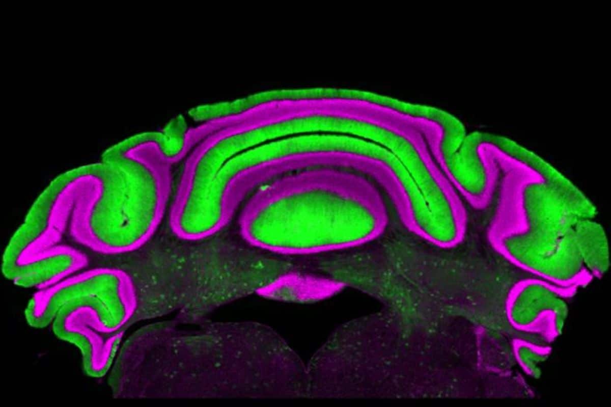
Image: The image is credited to Meike van der Heijden/Virginia Tech
Original Research: Open access.
“Steady-state Purkinje cell activity has limited predictive power for cerebellar output in disease” by Alyssa M Lyon, Viviana Hernandez-Castanon, Meike E van der Heijden. Journal of Physiology
DOI:10.1113/JP290000
Abstract
Steady-state Purkinje cell activity has limited predictive power for cerebellar output in disease
Cerebellar dysfunction can cause ataxia, dystonia, and tremor. Cerebellar nuclei neurons, the main cerebellar output neurons, exhibit distinct spike patterns in mouse models for different movement disorders. However nuclei spike pattern changes often arise from misfiring, miswiring, or degenerating Purkinje cells, which form the predominant input onto nuclei cells. It is often assumed that changes in Purkinje cell spike patterns cause inverse changes in nuclei cell spike patterns because Purkinje cells form GABAergic synapses onto cerebellar nuclei cells.
We test this hypothesis by investigating whether a systematic relationship between spike patterns in Purkinje and nuclei cells exists. We analysed parameters relating to spike rate and irregularity from single-cell in vivo electrophysiological recordings of cerebellar cells in five mouse models for cerebellar movement disorders. We examined whether changes in steady-state Purkinje cell spike patterns could predict nuclei cell spike pattern changes.
We found that some parameters for steady-state spike irregularity were positively correlated between Purkinje and nuclei cells, but no – especially no inverse – relationship was observed between steady-state Purkinje and nuclei cell spike rate.
We also did not observe an increased steady-state nuclei cell spike rate in mice with silenced or degenerating Purkinje cells. Because we find no systematic relationship between steady-state Purkinje and nuclei spike activity, it is not possible to reliably predict disease-associated nuclei spike patterns based on single-cell Purkinje recordings.
Moreover, normal Purkinje cell spike patterns can mask disease-causing spike patterns in cerebellar nuclei cells, underscoring the importance of studying cerebellar nuclei cell function in cerebellar disease.