Summary: Researchers constructed a high-resolution spatial map of the molecular changes induced by selective serotonin reuptake inhibitors (SSRIs) within the brain’s primary serotonin hub. Investigators utilized spatial transcriptomics to trace gene expression shifts over short- and long-term treatment with fluoxetine.
The findings upend the traditional view of the serotonin system as a uniform network, demonstrating instead that two distinct neuron populations respond in opposite directions to the same drug. This structural divergence perfectly mirrors the clinical timeline of antidepressant therapy, where transient side effects precede therapeutic relief.
Key Facts
- The Serotonin Homogeneity Fallacy: Antidepressants are globally ubiquitous, prescribed to more than 10% of the population in nations like Sweden, yet their exact impact on host gene expression inside serotonin neurons has remained poorly understood.
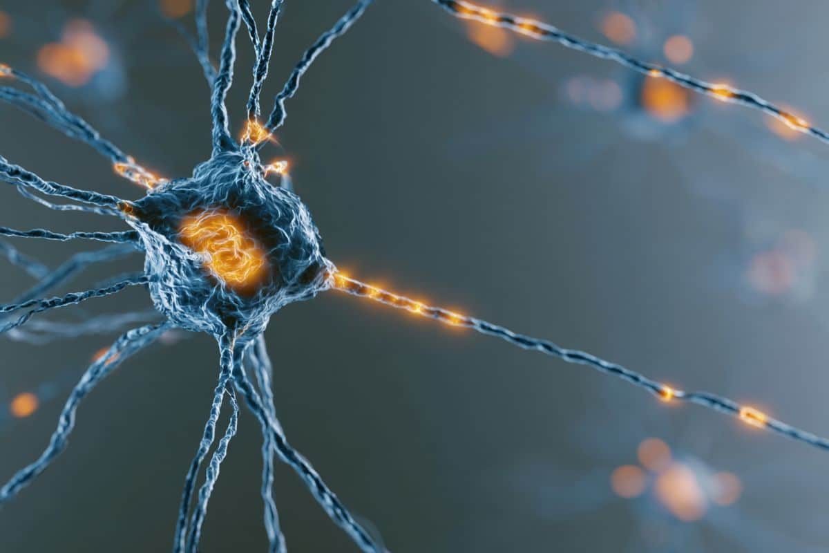
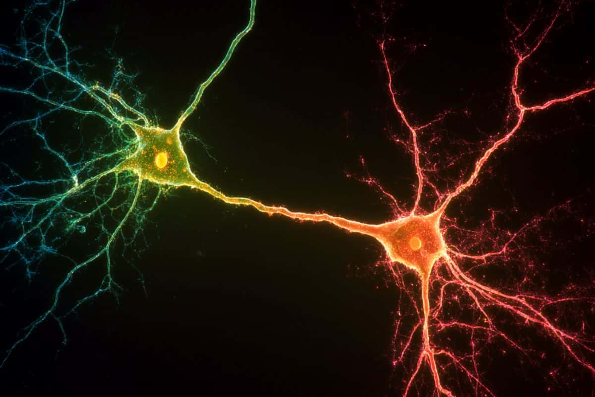
- High-Resolution Spatial Mapping: Researchers focused on fluoxetine, a widely prescribed SSRI, evaluating its molecular footprint inside the Dorsal Raphe Nucleus (the brain’s main serotonin-producing center). Spatial transcriptomics allowed the team to read out gene activity at single-cell resolution without blending distinct neuron types together.
- The Transient Phase (Group 1): Following short-term fluoxetine exposure, a specific subpopulation of serotonin neurons exhibited a sharp, temporary spike in the expression of the neuropeptide prodynorphin (Pdyn). Because Pdyn pathways are biologically tied to stress-induced depressive symptoms, this acute spike provides a concrete molecular explanation for the heightened anxiety and worsening mood patients frequently experience during their first days on an SSRI.
- The Delayed Therapeutic Phase (Group 2): Conversely, a separate subpopulation of serotonin cells responded exclusively to prolonged, chronic treatment by escalating the expression of thyrotropin-releasing hormone (TRH). TRH signaling is historically linked to anti-depressive functions, matching the multi-week delay required for clinical symptom relief to manifest.
- Next-Generation Drug Discovery: Lead investigator Dr. Iskra Pollak Dorocic emphasizes that identifying these opposite molecular candidate paths allows neuroscientists to bypass the unrefined “serotonin” label. These isolated pathways provide a direct blueprint to engineer targeted, next-generation antidepressants that preserve the late-stage TRH benefit while eliminating the initial Pdyn stress response.
Source: Stockholm University
Antidepressants are among the most widely prescribed medications in the world. In Sweden, more than one in ten people currently use an antidepressant, and SSRIs are by far the most common type.
“Yet we still understand surprisingly little about what these drugs actually do in the brain. Our study set out to map the gene-expression changes SSRIs induce in their primary target, the brain’s serotonin neurons”, says Iskra Pollak Dorocic, Assistant Professor at the Department of Biochemistry and Biophysics at Stockholm University.
Mapping changes
The study focused on fluoxetine, one of the most widely prescribed SSRIs, examining its effects on the brain’s main serotonin-producing region, the Dorsal Raphe Nucleus. Using a cutting-edge technique called spatial transcriptomics[LE1.1], the research group mapped changes in gene activity after both short-term and long-term treatment.
“Rather than treating the serotonin system as a single uniform population, we used spatial transcriptomics to read out gene activity at high resolution and map different types of serotonin neurons in the same brain area. That allowed us to see that these neurons are far more diverse than a single label suggests, and importantly that they do not all respond to the drug in the same way”, says Iskra Pollak Dorocic.
Two different paths
The study revealed widespread changes in gene expression following SSRI treatment. Most notably, the researchers identified two distinct subpopulations of serotonin neurons that responded differently to the drug:
- One group showed increased expression of the neuropeptide prodynorphin (Pdyn) after short-term treatment. Pdyn signaling has previously been linked to stress-induced depressive symptoms in other parts of the brain. However, this effect diminished with longer exposure to the antidepressant. The research suggests that this temporary increase in Pdyn could be linked to the negative effects that some patients experience when first starting SSRI treatment, such as increased anxiety or worsening mood.
- A second serotonin neuron population responded in the opposite way. These cells instead expressed the neuropeptide thyrotropin-releasing hormone (TRH), and their activity increased only after prolonged treatment. TRH signaling has previously been linked to anti-depressive functions in other parts of the brain. The findings suggest that TRH may play a role in the therapeutic effects of SSRIs that typically emerge after several weeks of treatment.
Good and bad effects
The discovery highlights the complexity of the brain’s serotonin system and suggests that different serotonin neurons may contribute to different phases of antidepressant response.
“We found that two distinct serotonin neuron populations are pushed in opposite directions by the same drug, one early and transiently, and one slowly over weeks. That mirrors the clinical picture, where unpleasant effects often come first and relief comes later, and it gives us concrete molecular candidates to interrogate next”, says Iskra Pollak Dorocic.
The genes, pathways and cell types identified in the study provide valuable leads for future research into the biological mechanisms underlying depression. The findings could also help guide the development of more targeted antidepressant treatments with fewer side effects and improved effectiveness.
Key Questions Answered:
A: It is driven by a temporary chemical spike in a specific group of brain cells. Stockholm University discovered that short-term SSRI exposure forces one population of serotonin neurons to rapidly produce a stress-linked neuropeptide called prodynorphin (Pdyn). This distressing reaction fades away as the drug stays in the system long-term.
A: Because the brain’s therapeutic pathways activate on a delayed, slower clock. A separate population of serotonin neurons increases its production of thyrotropin-releasing hormone (TRH), an anti-depressive signaling chemical, only after prolonged, chronic exposure to the medication.
A: By showing that “serotonin neurons” are not all identical. Instead of treating the whole system with a blanket drug, spatial transcriptomics maps different cell types in the exact same region. This allows chemists to design highly targeted drugs that can trigger the helpful TRH cells while entirely avoiding the anxious Pdyn cells.
Editorial Notes:
- This article was edited by a Neuroscience News editor.
- Journal paper reviewed in full.
- Additional context added by our staff.
About this neuroscience and SSRI research news
Author: Press Office
Source: Stockholm University
Contact: Press Office – Stockholm University
Image: The image is credited to Neuroscience News
Original Research: Open access.
“Effects of SSRIs on the spatial transcriptome of dorsal raphe serotonin neurons” by Charlotta Henningson, Jakub Mlost & Iskra Pollak Dorocic. Molecular Psychiatry
DOI:10.1038/s41380-026-03644-x
Abstract
Effects of SSRIs on the spatial transcriptome of dorsal raphe serotonin neurons
The serotonin system is the main therapeutic target for selective serotonin reuptake inhibitors (SSRIs) in treating depression, yet the mechanism of action of SSRIs remains incompletely understood. To investigate the molecular and transcriptional effects of SSRI administration on serotonin neurons, we performed spatial transcriptomics, a spatially resolved RNA-sequencing method in intact brain tissue.
Mouse brain sections containing the dorsal raphe nucleus and adjacent midbrain structures were analyzed, revealing six distinct serotonergic subpopulations with unique molecular signatures and spatial distributions. Both acute and chronic fluoxetine treatment induced a large number of changes in gene expression in the dorsal raphe nucleus.
Notably, Htr1a expression increased following acute treatment but decreased after chronic administration, supporting previous findings on serotonin transporter blockade effects on 5-HT1A autoreceptors. Gene enrichment and network analysis identified key pathways modulated by SSRI administration, including Ras, MAPK and cAMP signaling pathways as well as pathways involved in axonal guidance.
Additionally, we observed treatment-dependent opposing transcriptional changes in neuropeptides, particularly Thyrotropin-releasing hormone (Trh) and Prodynorphin (Pdyn), with distinct spatial localization within the dorsal raphe nucleus.
Collectively, our transcriptomic and in situ hybridization analyses reveal spatial and cell-type-specific heterogeneity in SSRI action within the dorsal raphe nucleus, providing new insights into the molecular basis of SSRI treatment effects.