Summary: Researchers identified a naturally occurring lipid molecule, erucamide, as a vital coordinator of the retina’s protective response to injury. The research demonstrates that while endogenous erucamide levels drop sharply as light-sensing photoreceptors degenerate, restoring the molecule stabilizes the retinal neurovascular unit. Rather than acting directly on photoreceptors, erucamide binds to the TMEM19 protein on CD11b⁺ myeloid immune cells.
This interaction activates the cells to release localized signals that preserve blood vessels and surrounding neural architecture. Delivered via engineered porous silicon nanoparticles to bypass its natural hydrophobicity, erucamide successfully slowed tissue breakdown in preclinical models, revealing a powerful new therapeutic pathway for progressive blinding diseases.
Key Facts
- Endogenous Signaling Decline: Erucamide is a naturally occurring fatty acid amide whose levels fall precipitously as photoreceptor cells die off during progressive retinal disease.
- The Environment-Targeting Shift: Instead of attempting to salvage dying photoreceptors directly, erucamide therapies work indirectly by optimizing the structural and immune health of the surrounding neurovascular environment.
- The TMEM19 Receptor Mechanism: Genetic or structural reduction of the newly identified binding protein TMEM19 completely halts erucamide’s protective efficacy, confirming it as the primary molecular gateway.
- Nanoparticle Delivery Breakthrough: Because erucamide is hydrophobic and prone to clumping in water-based mediums, the team utilized porous silicon nanoparticles to achieve stable, uniform distribution within the eye.
- Broad Therapeutic Scope: This approach targets foundational tissue degeneration mechanics, offering potential clinical interventions for diverse conditions including diabetic retinopathy, retinitis pigmentosa, and age-related macular degeneration.
Source: Scripps Research Institute
Many conditions that cause vision loss share a common feature: the gradual breakdown of the retina, the light-sensing tissue at the back of the eye. Although scientists know some of the structural changes that ensue as this damage progresses, less is understood about the molecular signals that shape how the retina copes with disease.
Now, a team at Scripps Research, in collaboration with UC San Diego and the Lowy Medical Research Institute, have found that a naturally occurring molecule called erucamide plays a role in how cells communicate in the retina.
Their study, published in Nature Neuroscience on June 19, 2026, found that while erucamide levels drop as light-sensing cells known as photoreceptors begin to die, restoring the molecule activates cellular responses that support retinal stability.
These findings suggest that erucamide may be part of a natural protective response in the retina, and could offer a new way to slow the progression of diseases that lead to vision loss.
“The retina doesn’t simply deteriorate; in fact, it actively responds to injury,” says senior author Martin Friedlander, a professor at Scripps Research. “Our work identifies erucamide as a signaling molecule that helps coordinate that response.”
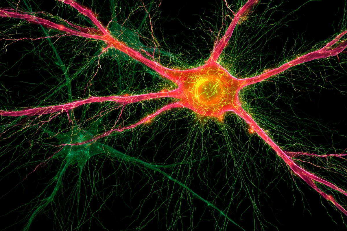
The retina depends on constant communication between neurons, glia, blood vessels and immune cells. This system, the neurovascular unit, keeps visual tissue functioning. But in diseases associated with loss of vision such as diabetic retinopathy, retinitis pigmentosa and age-related macular degeneration, that tight coordination wanes. Photoreceptors start dying, and eyesight fades.
The work from Friedlander’s team builds on earlier observations that transplanted stem cell-derived retinal cells could slow degeneration long after the cells had disappeared, suggesting they were releasing protective signals that outlasted their own survival. This insight prompted the team to search for the specific molecules responsible.
Though it’s long been recognized that fat-like compounds called lipids can act as signaling molecules in the body, many of these compounds haven’t been carefully studied in the context of retinal disease. To search for overlooked players, the scientists used mass spectrometry-based metabolomics: a technique that measures many small molecules in tissue at once. The team applied this approach to several well-established preclinical models of retinal degeneration, searching for molecules that changed as disease progressed.
Among the many molecules detected, erucamide stood out. Its levels fell sharply as photoreceptors began to deteriorate, suggesting the decline may not be incidental.
“That was a pivotal moment for us,” recalls co-author Dale Boger, the Richard and Alice Cramer Professor of Chemistry at Scripps Research. “It raised the possibility that erucamide could be influencing how tissue responds and wasn’t just changing as a consequence of disease.”
The team then set out to determine whether restoring erucamide could affect the retinal degenerative process. Using porous silicon nanoparticles—tiny, engineered delivery vehicles designed to release molecules in a controlled way—the scientists reintroduced erucamide into the eye. Because erucamide is hydrophobic (meaning it doesn’t dissolve well in water) and can form clumps when injected, the nanoparticles helped keep the molecule stable and evenly distributed.
Rather than acting directly on photoreceptors, erucamide activated a group of immune cells in the retina known as CD11b⁺ myeloid cells, which react to injury and support tissue health throughout the body. The team also identified a protein called TMEM19 that erucamide binds to; reducing TMEM19 levels prevented activation of these myeloid cells and blocked erucamide’s protective effects.
Once stimulated, myeloid cells released signals associated with neurovascular stabilization, supporting both the nerve cells and the blood vessels that nourish them. While the effect wasn’t an outright reversal of retinal damage, it slowed aspects of degeneration by preserving the structure and function of remaining tissue.
“Instead of targeting the photoreceptors themselves, erucamide appears to work by engaging the surrounding environment,” explains first author Guoqin Wei, a staff scientist at Scripps Research who began working on this project as a postdoctoral research associate in Friedlander’s lab seven years earlier. “That shift in perspective could be important for treating degenerative retinal diseases going forward.”
Although the team’s discovery offers a glimpse into how erucamide’s effects happen at the molecular level, additional studies are needed to clarify the full pathway. Future work will focus on erucamide signaling in various retinal diseases, and whether targeting this pathway can provide meaningful benefits over time.
And since erucamide’s hydrophobicity poses a formulation challenge—as most medicines for the eye are water-based—the scientists aim to improve how the molecule can be delivered as a therapy. They’re also planning to test modified versions of erucamide to see whether they produce stronger or more stable effects, while also investigating whether related lipid molecules may be even more effective than erucamide at activating protective responses.
Still, the findings highlight a broader notion: that some molecules already present in the body may be harnessed to support tissue under stress. The early results point to a potential strategy for slowing retinal degeneration by strengthening the tissue’s own response to damage.
“The goal is to reinforce a signal that’s already present,” notes Friedlander. “If we can learn how to modulate that response carefully, it could offer a new path for slowing the progression of retinal diseases where treatment options remain limited.”
Funding: This work was supported by funding from the Lowy Medical Research Institute; the National Eye Institute (grants R01EY11254 and 5R24EY017540); the California Institute for Regenerative Medicine (grant TR1-01219); the National Science Foundation through the UC San Diego Materials Research Science and Engineering Center (grant DMR-2011924); the National Institutes of Health (grants 2R01AI132413, R35 GM130385, U01 CA235493 and U01 CA305256); the National Institute on Drug Abuse (grant DA015648), the San Diego Nanotechnology Infrastructure of UC San Diego, a member of the National Nanotechnology Coordinated Infrastructure, which is supported by the National Science Foundation (grant ECCS-2025752); and the Natural Sciences and Engineering Research Council of Canada Postgraduate Scholarship–Doctoral program (grant NSERC PGS-D).
Key Questions Answered
A: The breakthrough relied on mass spectrometry-based metabolomics, an advanced analytic technique that maps hundreds of small lipid and metabolic molecules simultaneously within living tissue. By tracking several distinct preclinical models of retinal degeneration over an extended timeframe, the researchers isolated erucamide because its concentration curve dropped sharply in direct lockstep with the onset of photoreceptor cell death, indicating it was actively involved in the disease timeline.
A: Erucamide is highly hydrophobic, meaning it does not dissolve well in water. Because the interior environment of the eye and standard ophthalmic medications are primarily water-based, injecting raw erucamide causes the molecules to clump together into unstable, ineffective aggregates. Enclosing the lipid inside porous silicon nanoparticles keeps the compound perfectly stable, preventing clumping and allowing a slow, controlled, and uniform release across the damaged retinal layers.
A: Traditional therapies try to preserve dying photoreceptors directly, which is exceptionally difficult once the surrounding support systems break down. Erucamide shifts the perspective by targeting CD11b⁺ myeloid immune cells instead. Once activated via the TMEM19 protein, these helper cells naturally repair and stabilize the entire neurovascular unit, the delicate web of blood vessels and helper cells that feed the retina. By restoring the environment first, the remaining visual cells are given the baseline structural support they need to survive.
Editorial Notes:
- This article was edited by a Neuroscience News editor.
- Journal paper reviewed in full.
- Additional context added by our staff.
About this visual neuroscience research news
Author: Press Office
Source: Scripps Research
Contact: Press Office – Scripps Research
Image: The image is credited to Neuroscience News
Original Research: Open access.
“A fatty acid amide activates myeloid cells and improves neurovascular outcomes in retinal degeneration” by Guoqin Wei, Shreyosree Chatterjee, Qinglin Yang, Sanahan Vijayakumar, Daisuke Ogasawara, Sarah Giles, Katie Biscocho, Peter Westenskow, Junhua Wang, Ruhan Fan, Helena Pham, Edith Aguilar, Jacob Robinson, Ayumi Usui-Ouchi, Roberto Bonelli, Kevin Eade, Gary Siuzdak, Benjamin Cravatt, Michael J. Sailor, Dale Boger & Martin Friedlander. Nature Neuroscience
DOI:10.1038/s41593-026-02341-w
Abstract
A fatty acid amide activates myeloid cells and improves neurovascular outcomes in retinal degeneration
Neurovasculoglial cross-talk underlying breakdown of the neurovascular unit is a central, yet poorly understood, component of many neurodegenerative disorders of the CNS, including retinal disease. Primary fatty acid amides have been identified to regulate this cross-talk between vasculature and neuronal tissues, but specific molecules and mechanisms remain unresolved.
Here we show, using an unbiased high-resolution metabolomics screen, that erucamide, a 22:1 monounsaturated omega-9 fatty acid amide, is highly dysregulated during photoreceptor degeneration in mice. In vivo delivery of erucamide using organosilane-modified porous silicon nanoparticles activated retinal myeloid cells, leading to the upregulation of angiogenic and neurotrophic cytokines that limited vascular and neuronal degeneration.
We identified TMEM19 as a binding protein for erucamide that is crucial for myeloid cell activation and subsequent neuroprotection.
These findings reveal a previously unknown primary fatty acid amide pathway that modulates neuroimmune interactions during retinal degenerative diseases. We propose erucamide and analogs as candidate therapeutics.